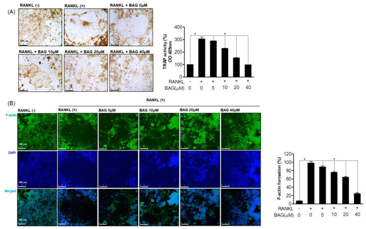
Figure 2.
Inhibitory Effect of BAG on RANKL induced osteoclast differentiation. (A) RAW264.7 cells were seeded in a 24 well plate at a density of 5 x 10³ and treated with RANKL with various concentrations of BAG (5–40 μM) for 5 days, and then TRAP staining and activity were measured. (B) As a representative fluorescence micrograph of the effect of BAG on F-actin belt formation, the indicated concentrations of BAG (5–40 μM) were treated with RANKL for 5 days. The actin cytoskeleton was then stained with Alexa 488-Phalloidin (green), and the nuclei were counterstained with DAPI (blue). * p < 0.05, versus the RANKL treated group.

