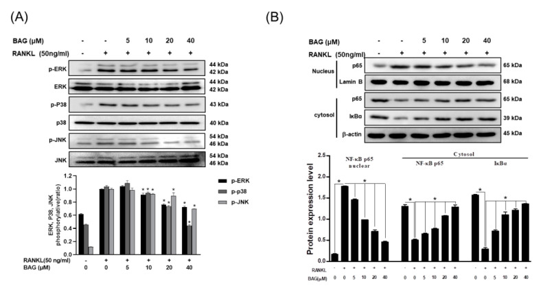
Figure 6.
BAG suppresses RANKL-induced activation of NF-kβ and MAPKs. (A) RAW264.7 cells (5 × 10⁵ cell/mL) were seeded for 24 h, then pre-incubated with the indicated concentration of BAG for 1 h and stimulated with 50 ng/mL of RANKL for 30 min and were subjected to Western blot analyses using specific antibodies’ phosphorylated forms of ERK, p38, and JNK. (B) NE-PER (Nuclear and Cytoplasmic Extraction Reagents) kits were used for separation of cytoplasm and nucleus, and Western blot analysis was performed on each cytoplasm and nucleus. Graphs show normalized expression of each indicated protein against the expression of β-actin or lamin B. * p < 0.05 versus the RANKL-treated group.

