Figure 2.
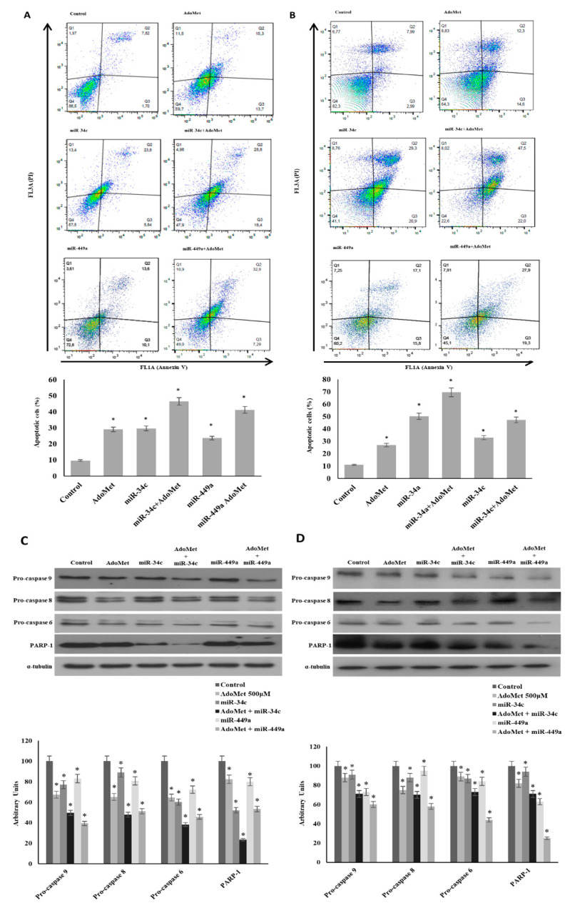
Effect of AdoMet/miR-34c and AdoMet/miR-449 combination on apoptosis and levels of some relevant apoptosis-related proteins in MDA-MB-231 and MDA-MB 468 cells. MDA-MB 231 and MDA-MB 468 cells were transfected with 100 nM miR-34c or miR-449a mimic supplemented or not (Control) with 500 μM AdoMet for 72 h. Apoptosis of MDA-MB 231 (A) and MDA-MB 468 cells (B) was evaluated by FACS analysis. Representative dot plots of both Annexin V-FITC and propidium iodide (PI)-stained cells are shown. The different quadrants report the percentage of cells: Viable cells, lower left (Q4); early apoptotic cells, bottom right (Q3); late apoptotic cells, top right (Q2); and non-viable necrotic cells, upper left (Q1). For each sample 2 × 10⁴ events were acquired. The analysis was carried out by triplicate determination of at least 3 separate experiments. The lower left and lower right histogram plots show the percentage of apoptotic cells, respectively, for a single treatment. * p < 0.05 versus untreated cells (Control). The expression levels of pro-caspase 9, pro-caspase 8, pro-caspase 6, and PARP-1 were detected by Western blot analysis using the total cell lysates of MDA-MB-231 (C) and MDA-MB-468 (D). The densitometric analysis was reported. Data are reported as percentage of protein expression of untreated control (100%). The house-keeping protein α-tubulin was used as loading control. The images are representative of three immunoblotting analyses obtained from at least three independent experiments. Uncropped images of Western blots are reported in Figure S1.

