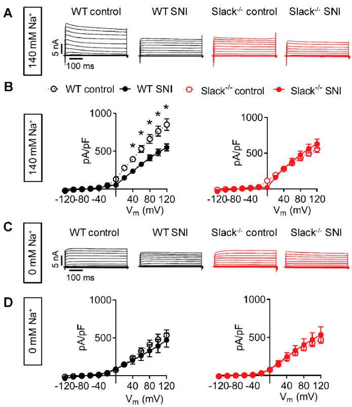
Figure 2.
Slack-mediated potassium currents in sensory neurons are reduced after SNI. (A,B) Representative outward K+ current (IK) traces (A) and associated current-voltage (I-V) curves (B) from whole-cell voltage recordings on IB4-positive lumbar (L4-L5) dorsal root ganglion (DRG) neurons of WT (black) and Slack-/- mice (red) 14–19 days after spared nerve injury (SNI). Contralateral DRG neurons were used as control. Recordings shown in (A) and (B) were performed in the presence of 140 mM NaCl in the external solution, i.e., under physiological conditions. n = 21–29 cells per group. Repeated ANOVA measures followed by Fisher’s Least Significant Difference test; WT control versus WT SNI: p = 0.0062; Slack-/- control versus Slack-/- SNI: p = 0.4374. (C,D) Representative IK traces (C) and associated I-V curves (D) in the same experimental setting as shown in (A) and (B), however, after replacement of NaCl by 140 mM choline chloride in the external solution to obtain Na+-free conditions. n = 7–9 cells per group. Repeated ANOVA measures: WT control versus WT SNI: p = 0.1825; Slack-/- control versus Slack-/- SNI: p = 0.6125. The data show that Na+-activated IK (IKNa) in sensory neurons is carried by Slack channels and reduced after SNI. Data in (B) and (D) are mean ± SEM. * p ˂ 0.05.

