Figure 4.
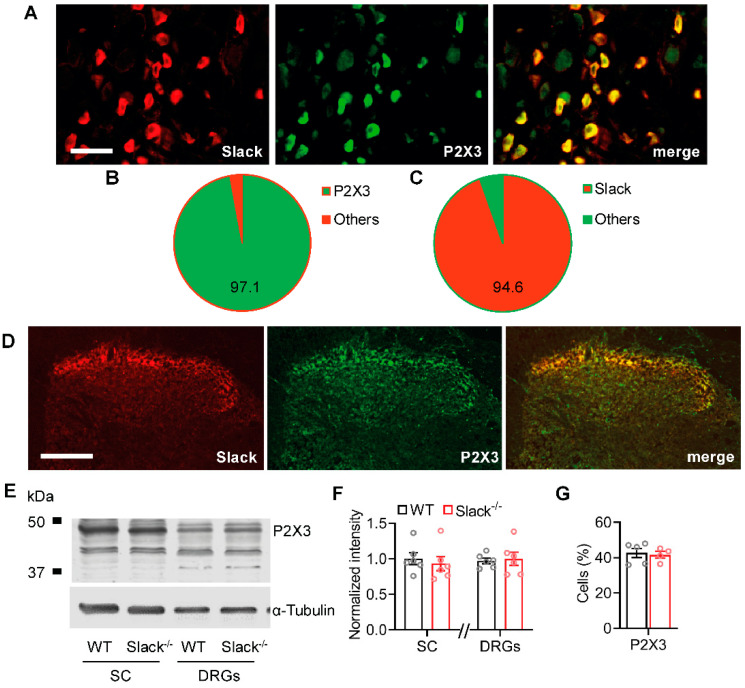
Slack channels co-localize with P2X3 receptors in sensory neurons. (A–C) Double-labeling immunostaining of Slack and P2X3 in sensory neurons (A) revealed that 97.1% ± 0.2% of Slack-positive DRG neurons co-stained with P2X3 ((B); 1203 cells counted, n = 3 mice) and that 94.6% ± 2.0% of P2X3-positive DRG neurons co-stained with Slack ((C); 1203 cells counted, n = 3 mice). (D) Double-labeling immunostaining of Slack and P2X3 in the spinal cord indicates a high degree of co-localization in the superficial dorsal horn. (E,F) Western blot of P2X3 in spinal cord (SC) and DRGs from WT and Slack-/- mice demonstrates identical abundance of P2X3 in both genotypes. The uncropped original image is shown in Figure S2B. Student’s t-test: p = 0.5986 in the spinal cord and p = 0.7631 in DRGs. Alpha-tubulin was used as a loading control. (G) Immunostaining revealed that the percentage of DRG neurons positive for P2X3 is similar in WT and Slack-/- mice. Student’s t-test: p = 0.4046. Bars denote mean ± SEM. Scale bars: 50 µm (A) and 100 µm (D).

