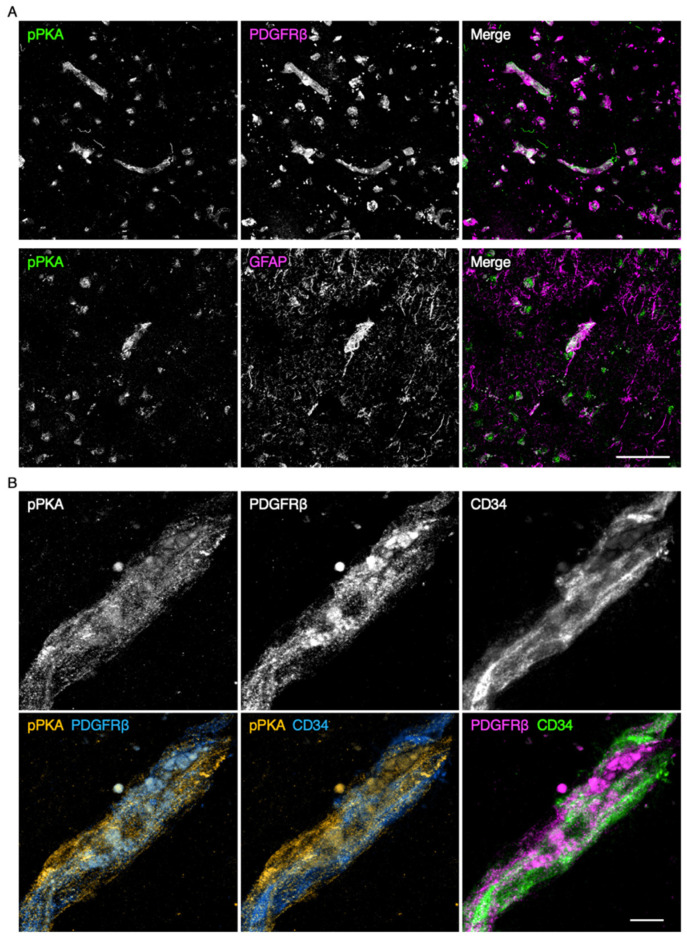
Figure 3.
Protein kinase A (PKA) activation in the microvascular endothelial cells and pericytes of the schizophrenic PFC. (A) Confocal images of the schizophrenic PFC gray matter stained for phospho-PKA (pPKA) and either PDGFRβ or glial fibrillary acidic protein (GFAP). (B) Confocal images of the schizophrenic PFC gray matter stained for pPKA, PDGFRβ, and CD34. Scale bars, 50 µm (upper); 10 µm (lower).

