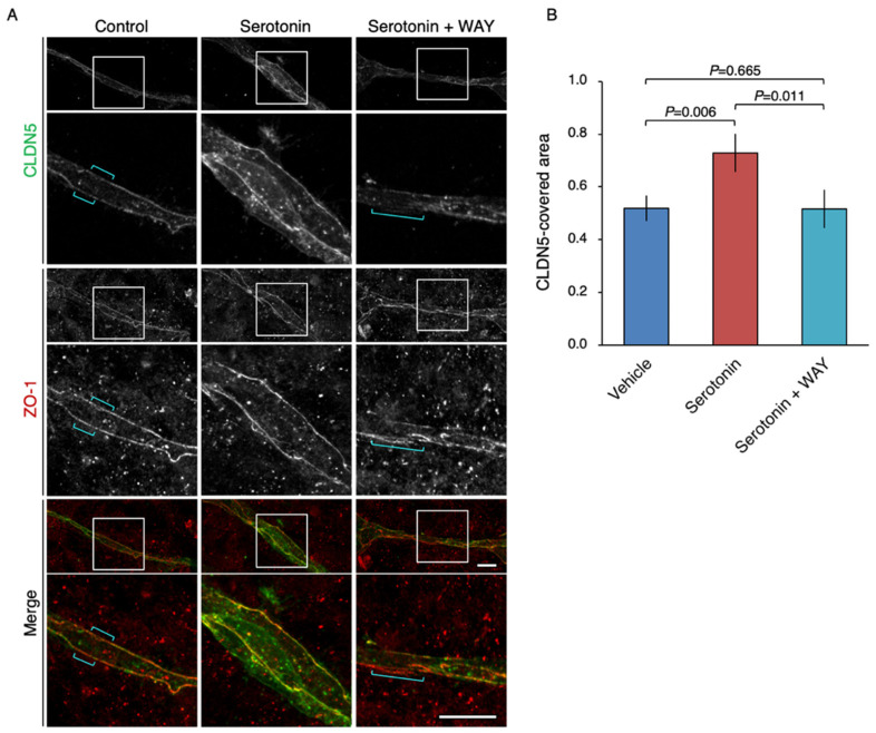
Figure 6.
Up-regulation of the CLDN5-immunoreactive area in the microvascular endothelial tube-like structure via the serotonin/5-HT1A receptor signaling. (A) Confocal images of two-dimensional co-culture stained for CLDN5 and ZO-1. The HBPCT and the human primary BMVECs were grown under two-dimensional co-culture conditions for 4 days. Brackets indicate the breakdown of CLDN5. WAY: WAY-100635. Scale bars, 50 µm. (B) The CLDN5-length is divided by the ZO-1-length, and the relative CLDN5-covered area is shown in histograms (mean ± SD; n = 3). Similar results were obtained from three independent experiments.

