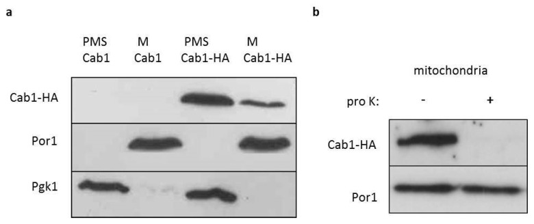
Figure 1.
Localization of Cab1p. (a) Equal amounts (20 mg) of the mitochondrial fraction (M) and post-mitochondrial supernatant (PMS) extracted from Δcab1 strain harboring pFL39CAB1 or pFL39CAB1HA were resolved by SDS-PAGE and analyzed by immunoblotting with HA, Pgk1 (cytosolic marker) and Por1 (mitochondrial outer membrane marker) antibodies. The experiment was performed on two independent clones for each strain (N = 1; n = 2) (b) Mitochondria were treated at 4 °C for 60 min with proteinase K (pro K) (1 mg/mL). The filter was incubated with anti-HA and anti-Por1 antibodies. The experiment was performed on two independent clones of Δcab1 strain harboring pFL39CAB1HA (N = 1; n = 2).

