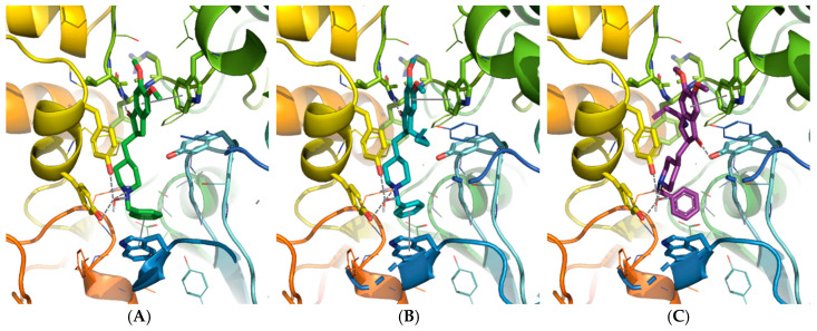
Figure 3.
Donepezil position from the X-ray structure (A) compared to the enantiomer R,R (B) and S,S (C) of trans-19 positioned in (h)AChE binding sites using the docking studies. The compounds and the selected side chains of the binding site residues are in stick and the protein in ribbon representation. This figure was made with PYMOL (DeLano Scientific, 2002, San Carlo, CA, USA).

