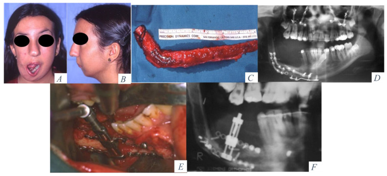
Figure 3.
(A) Hemimandibular agenesis with facial asymmetry and mandibular deviation. (B) Retrusion of the lower facial third. (C) Mandibular reconstruction with an osseous fibula flap. (D) Panoramic radiograph with vertical discrepancy between the remaining mandible and the fibula flap. Le Fort I osteotomy for occlusal compensation. (E) Alveolar distractor placed in the fibula. (F) Panoramic radiograph at the beginning of the distraction procedure.

