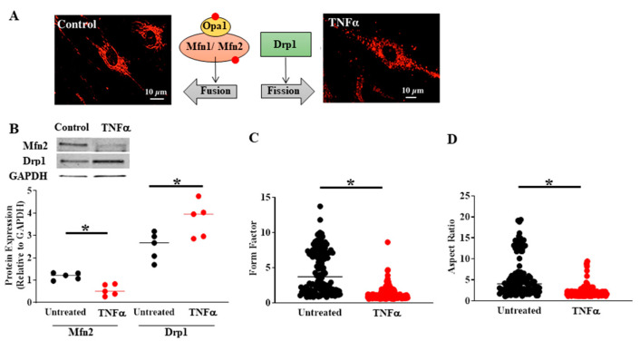
Figure 4.
Compared to untreated controls, TNFα (20 ng/mL, 24 h) exposure induced mitochondrial fragmentation in human ASM (A) associated with reduced Mfn2 and increased Drp1 protein levels (B). The extent of mitochondrial fragmentation was assessed by calculating (C) form factor and (D) aspect ratio of mitochondria labeled using MitoTracker Red (A). In B, cells were dissociated from ASM samples from n = 5 patients and divided into TNFα treated and untreated control groups. In C and D, multiple mitochondria/cells were measured from ASM cells isolated from n = 5 patients divided into TNFα treated and untreated control groups. (* denotes a significant difference between TNFα treated and untreated control groups; p < 0.05; t-test). All data are presented as scatter plots with lines indicating mean values. These figures are modified from presentations of previously reported results [4].

