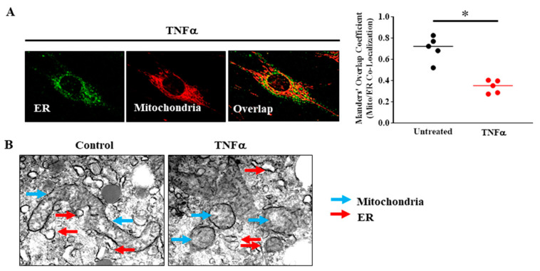
Figure 5.
(A) In human ASM cells, ER was labeled using BODIPY FL thapsigargin (endoplasmic reticulum Ca2+ pumps, green) and mitochondria were labeled using MitoTracker red and imaged in 3D by confocal microscopy. Proximity of mitochondria to the ER was determined by measuring the Manders’ overlap co-efficient. Acute TNFα exposure (20 ng/mL, 24 h) reduced proximity of mitochondria to the ER (* denotes a significant difference between TNFα treated and untreated control groups; p < 0.05; t-test; cells were dissociated from ASM samples from n = 5 patients and divided into TNFα treated and untreated control groups). (B) 3D EM images showing that mitochondria (blue arrows) in human ASM cells are fragmented after TNFα (20 ng/mL, 24 h) exposure and that the incidence of close proximity of mitochondria to ER (red arrows) was reduced (cells were dissociated from ASM samples from n = 2 patients and divided into TNFα treated and untreated control groups). All data are presented as scatter plots with lines indicating the mean. These figures are modified from presentations of previously reported results [13].

