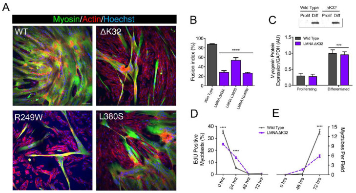
Figure 1.
In vitro myoblast fusion and myotube formation. (A) Confocal immunofluorescence images of myosin (MF20, green) in wild-type (WT) and LMNA-related congenital muscular dystrophy (LMNA-CMD) mutant (ΔK32, L380S and R249W) cells, after 3 days of differentiation. Nuclei are stained with Hoechst (blue). Scale bar = 100 µm. (B). Fusion index in WT and LMNA-CMD mutant cells after 3 days of differentiation. Pooled values of WT (WT1 and WT2) are presented. n = 10 fields of view analyzed per time point from across 3 separate experiments. Values are expressed as means ± SEM. **** p < 0.0001 versus WT myotubes. (C) Myogenin expression in WT and LMNA ΔK32 mutant cells in proliferation and after 3 days of differentiation. n ≥ 3 from at least 2 separate experiments. *** p < 0.001 versus WT myotubes. (D) EdU positive myoblasts (%) and (E) number of myotubes per field until 3 days of differentiation. n = 10 fields of view analyzed per time point from across 3 separate experiments. Values are expressed as means ± SEM. **** p < 0.0001 versus WT cells.

