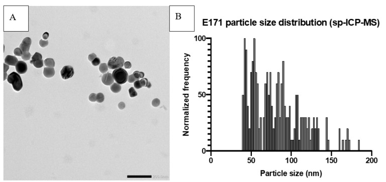
Figure 1.
Example of E171 particle characterization. Prior analysis the samples were dispersed according to the NanoGenotox dispersion protocol at a final concentration of 2.56 mg/mL in 0.05% BSA solution and probe sonicated on ice for 16 min (4 W). (A) Transmission Electron Microscope picture of E171. (B) Size distribution of E171 particles, measured by single-particle ICP-MS, with a median particle size of 79 nm and 72% of particles < 100 nm.

