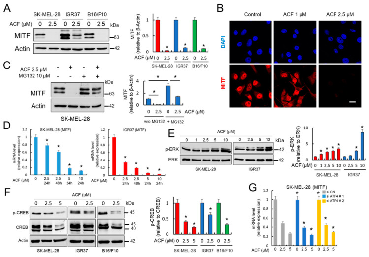
Figure 3.
ACF decreases MITF expression in melanoma cells. (A) Effect of ACF (24 h) on MITF expression in melanoma cells analyzed by western blot. IOD quantification is shown (histogram; * p < 0.05 when compared with untreated controls). (B) Confocal microscopy analysis (63X magnification) of MITF in SK-MEL-28 melanoma cells under indicated conditions (cells were treated with ACF for 24 h). Bars, 27 μM. (C) Western blot experiments for the effect of ACF on MITF expression in the absence or the presence of MG132. SK-MEL-28 cells were treated for 12 h with ACF alone or simultaneously co-treated with ACF and MG132. * p < 0.05 when comparing indicated data groups. (D) qRT-PCR analysis of MITF mRNA in indicated melanoma cells before and after ACF treatments (24 h). Relative mRNA expression in treated cells was normalized with respect to untreated cells. * p < 0.05. (E) Effect of ACF (24 h) on the phosphorylation of ERK1/2 (p-ERK) in melanoma cells analyzed by western blot. Specific antibodies recognized the diphosphorylated forms of ERK1/2 (Thr183 and Tyr185 based in ERK2 nomenclature). Constitutive total ERK was used as a reference for p-ERK expression. IOD quantification is shown (histogram; * p < 0.05 when compared with untreated controls). (F) Effect of ACF (24 h) on Ser133 phosphorylation in CREB (p-CREB). Western blot of total CREB showed two clear bands at 40 and 45 kDa, corresponding to the upper band to phosphorylated CREB. IOD quantification is shown (histogram; * p < 0.05 when compared with untreated controls). (G) Effective silencing of ATF4 with two different siRNAs (Figure 2F) did not influence MITF-mRNA expression in SK-MEL-28 cells. * not significant when compared to siATF4 samples with their respective treatments in siCN samples. When indicated, cells were treated with ACF for 24 h.

