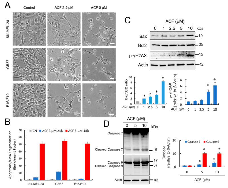
Figure 5.
ACF induces apoptotic cell death in melanoma cells. (A) Morphological aspect of untreated melanoma cells compared with those subjected to 2-days of treatment with indicated concentrations of ACF (bars, 100 μM). 40× magnification (B) Apoptosis determination at different ACF concentrations in indicated melanoma cells after 24 and 48 h of treatment. Data were obtained in triplicate in two independent experiments. Differences in apoptosis in ACF-treated cells were significant with respect to untreated controls for each drug concentration and at any time (p < 0.05). (C) Western blots showing the effect of ACF on Bax, Bcl2, and p-γH2AX proteins. SK-MEL-28 cells were treated with different concentrations of ACF for two days. The ratios between Bax and Bcl2 and relative p-γH2AX are presented in the histograms (* p < 0.05 when compared with the untreated control). (D) Western blot analysis of caspase 7 and caspase 9 in the control SK-MEL-28 cells and those treated with indicated doses of ACF. IOD quantification is shown (histogram; * p < 0.05 when compared with their respective untreated controls).

