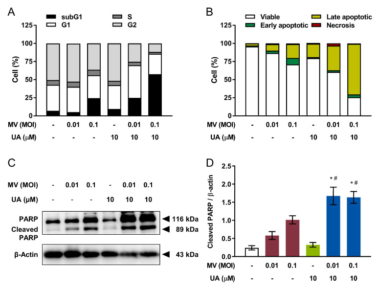
Figure 3.
Co-treatment using UA and oncolytic MV enhances apoptotic cell death in human breast cancer MCF-7 cells. MCF-7 cells were first treated with combination of UA (10 µM) and MV (MOI 0.01 or 0.1) for 5 days, then analyzed by flow cytometry for (A) cell cycle distribution and (B) apoptosis induction, using propidium iodide (PI) staining and double staining (PI and Annexin V conjugated with allophycocyanin [APC]) respectively. Percentages shown are determined by Beckman Cytomics TM FC500 Flow Cytometry CXP analysis software. (C) Lysates of MCF-7 cells co-treated with UA (10 µM) and MV (MOI 0.01 or 0.1) for 5 days were analyzed by western blot for poly (ADP-ribose) polymerase (PARP) cleavage. (D) Quantitative analysis of the relative level of cleaved PARP from (C). All quantitative data are expressed as means ± SEM from three independent experiments; * p < 0.05 compared with MV MOI 0.01 or 0.1, # p < 0.05 compared with UA treatment only.

