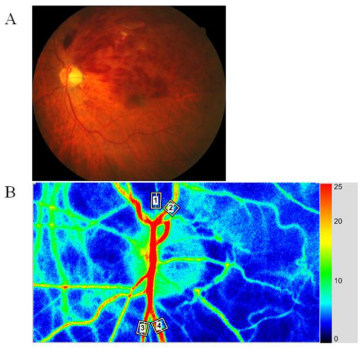
Figure 1.
Representative fundus color photograph, and representative relative blood volume data obtained with laser speckle flowgraphy (LSFG). (A) Fundus color photograph shows branch retinal vein occlusion (BRVO). The upper part is the occluded region, and the lower part is the non-occluded region. (B) Blood flow was automatically tracked in an artery (white square #1) and vein (white square #2) in the occluded region and in an artery (white square #3) and vein (white square #4) in the non-occluded region.

