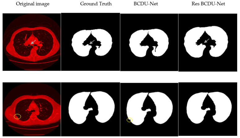
Figure 17.
Visualizes the challenges for segmentation. First row presents the challenge of considering micro pulmonary tissues in the segmented image as the non-pulmonary region causing high false positive. Second row presents the challenge of losing attached nodules to the lung wall. (A yellow circle wrapped around the center of the nodule).

