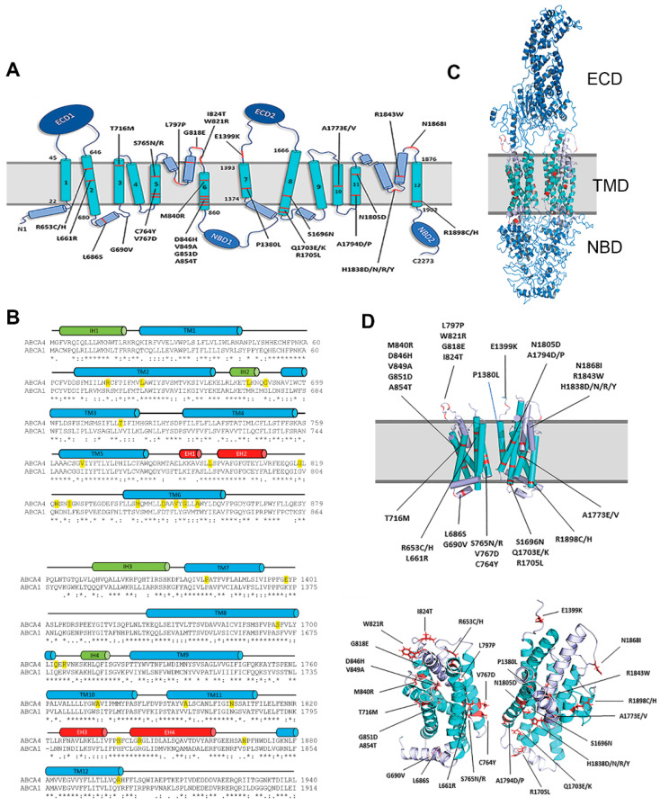
Figure 1.
Location of TMD missense mutations in ABCA4 associated with STGD1. (A) Topological model of ABCA4 showing the location of mutations in TMD1 and TMD2. ECD—Exocytoplasmic Domain; TMD—Transmembrane Domain; NBD—Nucleotide Binding Domain. (B) Sequence alignment of the Transmembrane Domains TMD1 and TMD2 of ABCA4 and ABCA1 using Clustal Omega. The locations of the α-helical membrane spanning segments are shown as blue cylinders (TM1-12), intracellular transverse coupling helices are in green (IH1–IH4), and exoplasmic V-shaped α-helical hairpin helices are in red (EH1–EH4). The STGD1 missense variants examined in this study are highlighted in yellow. (C) Homology model of ABCA4 based on the cryoEM structure of ABCA1. (D) The location of disease-causing mutations within the TMD1 and TMD2. Top: Transverse view relative to the membrane with the transmembrane segments shown as cylinders. Bottom: Top view relative to the membrane with the transmembrane segments shown as ribbons.

