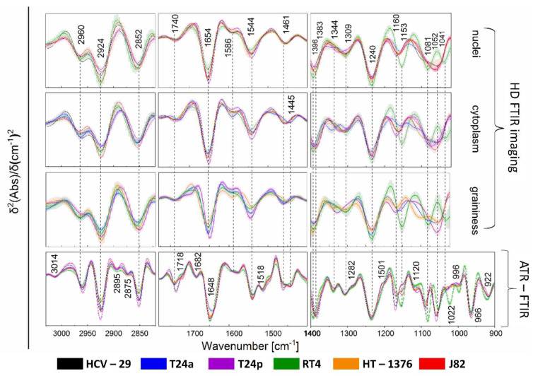
Figure 3.
Averaged second derivatives of FTIR transmission spectra of the nuclei and cytoplasm for each cell line and graininess observed for T24a, T24p, RT4 and HT1376 cells compared to ATR−FTIR spectra of whole cells. Gray shading denotes standard deviation (±SD); N = 20 spectra per cell line. Averaged normal FTIR spectra are shown in Figure S3 in SM.

