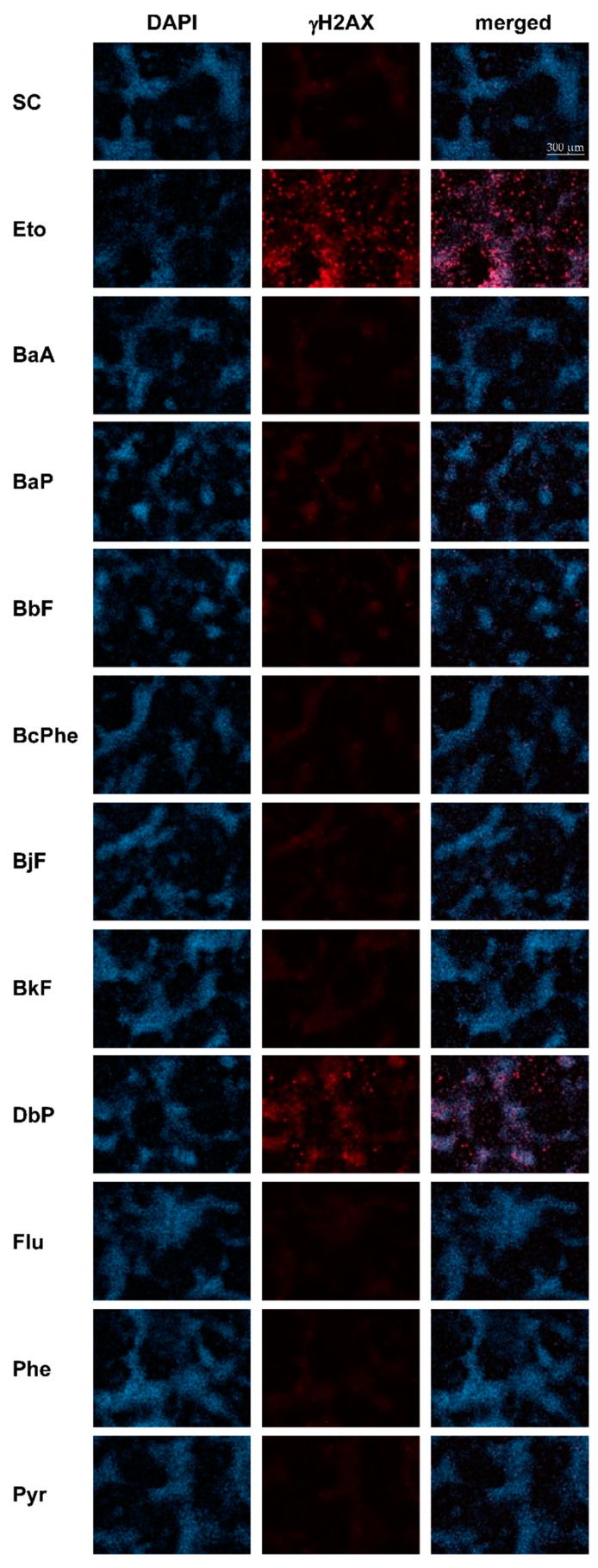
Figure 5.
Frequency of histone H2AX phosphorylation. HepaRG cells were treated with 1, 5, or 10 µM of each PAH and incubated for 12, 24, 48 h. Etoposide (Eto; 25 µM) served as a metabolism-independent positive control. Afterward, cells were fixed, blocked, and stained with a combination of primary anti-γH2AX and secondary Alexa Fluor 647-antibody (red). Nuclei were counterstained using 4′,6-diamidino-2-phenylindole (DAPI) (blue). The figure depicts representative fluorescence images (brightness +30%) of each channel as well as a merged picture, respectively, 48 h posttreatment with solvent control (SC), Eto, or 10 µM PAH. Images were taken with Celldiscoverer (Zeiss, Oberkochen, Germany) and Zeiss Blue software (Zen 3.1 blue edition, Zeiss, Oberkochen, Germany).

