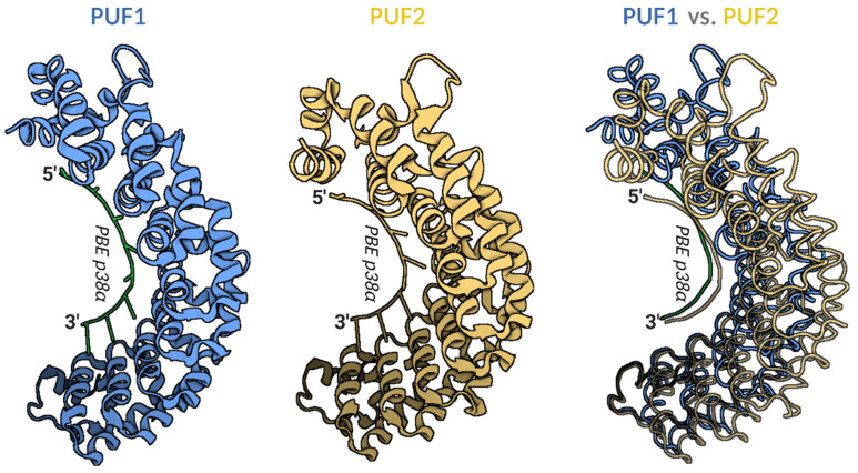
Figure 1.
Models of human PUF1 and PUF2 RNA-binding domain crystal structure in complex with PUM-binding element (PBE) of p38α mRNA target. Left panel—cartoon model of PUF1 structure in a complex with PBE of p38α mRNA (PDB id:3Q0M); middle panel—cartoon model of PUF2 structure in a complex with PBE of p38α mRNA (PDB id:3Q0R); right panel—overlay of tube models of PUF1 (blue) and PUF2 (yellow) structures in complex with PBE of p38α mRNA. The overlay panel shows a more open PUF1 compared to the PUF2-domain, binding the same PBE of p38α mRNA. Visualized models were obtained using BioRender.com from 3Q0M and 3Q0R PDB structures [10]. PUF—PUM RNA-binding domain; PBE—PUM binding element UGUAHAUW; PDB—Protein Database Bank.

