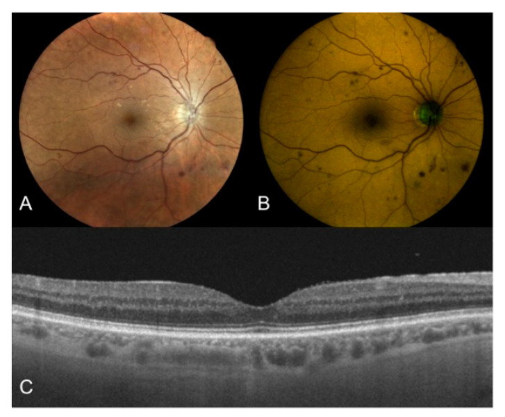
Figure 2.
Right eye of a 68-year old male patient affected by type 2 DM with moderate DR. (A) True-color fundus photography of the posterior pole showing multiple hemorrhages and initial vitreo-retinal interface syndrome; (B) Color-FAF of the same eye with detected values of foveal GEFC and REFC intensity of 17 and 26, respectively; (C) OCT horizontal B-scan centred on the fovea showing a dry macula with normal reflectivity of the inner and outer retinal layers. DM: diabetes mellitus; DR: diabetic retinopathy; FAF: fundus autofluorescence; GEFC/REFC: green/red emission fluorescence components; OCT: optical coherence tomography.

