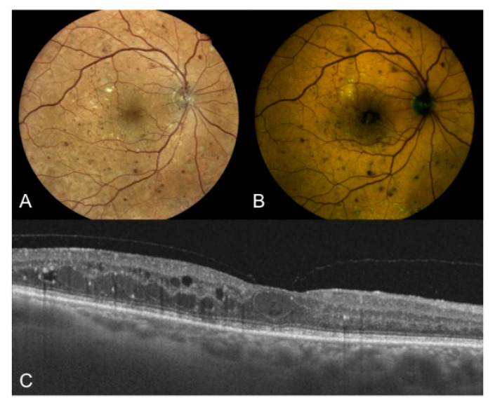
Figure 3.
Right eye of a 67-year old male patient affected by type two DM with proliferative DR. (A) True-color fundus photography of the posterior pole showing NVD, multiple hemorrhages, extrafoveal hard exudates, cystoid macular edema, and peripheral laser treatment; (B) color-FAF of the same eye with detected values of foveal GEFC and REFC intensity of 31 and 40, respectively; (C) OCT horizontal B-scan centered on the fovea showing center involving cystoid macular edema (CRT = 356 μm), hard exudates, and hyper-reflective intraretinal spots. DM: diabetes mellitus; DR: diabetic retinopathy; NVD: new vessels at disc; FAF: fundus autofluorescence; GEFC/REFC: green/red emission fluorescence components; OCT: optical coherence tomography; CRT: central retinal thickness.

