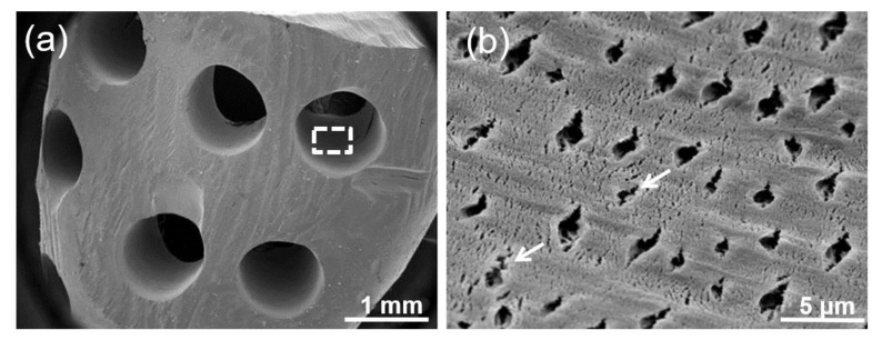
Figure 3.
(a) Scanning electronic microscopy (SEM) image of PR-DDM showing smooth demineralized dentin surface with custom-made artificial macro-pores. (b) Higher magnification of white dotted line in Figure a showing exposed dentinal tubule with micro-cracks (white arrow) in a dense collagen matrix.

