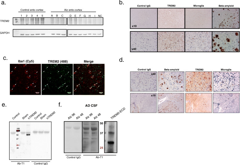
Fig. 2.
Recognition of TREM2 in AD models/human by Ab-T1. a Western blot showing Ab-T1 binding to human entorhinal cortex extracts from Alzheimer/control group patients. HEK293T protein lysate was used as negative control (NC). GAPDH as “housekeeping” protein loading control is shown in lower panels. b Immunohistochemistry staining showing Ab-T1 binding to human brain sample (entorhinal cortex sections) from Alzheimer’s disease patient (TREM2) with microglia and beta amyloid staining of same human brain tissue sample. Mouse IgG was used as negative control staining. c Confocal microscope scan images showing co-localization of TREM2 (mouse Ab-T1) with resident Iba1 positive cells (Microglia) in 5xFAD mice brain slices (white arrows). d Immunohistochemistry staining showing Ab-T1 binding to brain tissue from 5xFAD mice (TREM2) with microglia and beta amyloid staining of same mice tissue sample. Mouse IgG was used as negative control. e Western blots of supernatants (left panel) for soluble TREM2 detection in parental HEK293T vs. HEK293T-hTREM2 cells using mouse Ab-T1 and mouse IgG1 as control Ab. Transfected HEK293T cells with no insert DNA vector were used as sham. f Western blots of soluble hTREM2 detection in human CSF from Alzheimer patients using Ab-T1 and mouse IgG1 as control IgG antibody. TREM-ECD represents soluble TREM2 recombinant protein control

