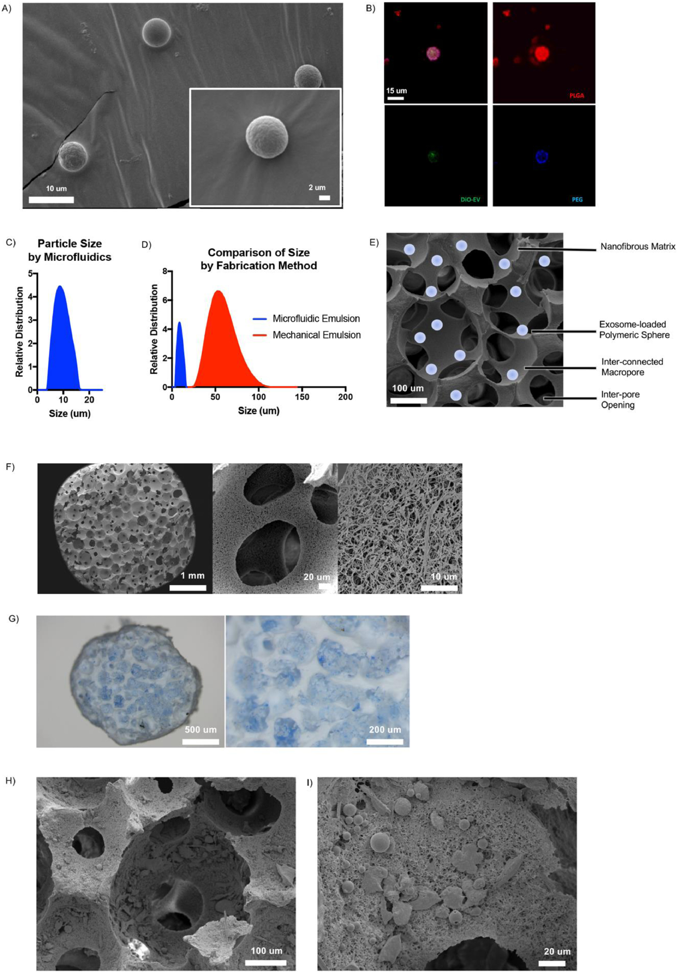
Fig 5.

Characterization of EXO-MS. Scanning electron microscopy images of EXO-MS prepared by microfluidic double emulsion method showed smooth, uniformly sized spheres (A). EXO-MS made from PLGA-PEG-PLGA block copolymer were mixed with fluorescently labelled polymer blocks and visualized by confocal microscopy; PEG (blue) segments self-assembled on the inside of the particle to form a sphere wherein OS-EXO (green) localize, among a disperse PLGA (red) microspheres (B; scale: 15 um). The size of EXO-MS was measured by a laser diffraction method (C, davg = 7.70 um). Compared to self-assembling particles from the same polymer made under mechanical emulsion conditions, particles made by a microfluidic platform were significantly smaller and less disperse (D). EXO-MS were attached to poly(l-lactic acid) (PLLA) tissue engineering scaffolds, as shown in the schematic (E; scale: 100 um). Scaffolds were a matrix of interconnected macropores with a nanofibrous surface texture (F). EXO-MS were seeded onto the pore surface of the scaffold using a solvent-wetting method, visualized by light microscopy (G, dyed blue for visual contrast) and scanning electron microscopy (H, I).
