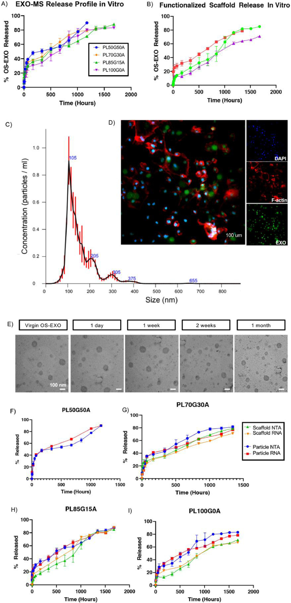
Fig. 6.

Evaluation of the controlled release of OS-EXO from EXO-MS. The release profile of OS-EXO was modulated by the hydrophilicity of the copolymer (A). After attachment of EXO-MS to PLLA scaffolds, the release of OS-EXO became more linear (B). OS-EXO released from EXO-MS at 2 weeks maintained their characteristic size (davg = 105 nm), evaluated by nanoparticle tracking analysis (C). Primary mBMSCs were treated with EXO-MS eluent (collected at 2 weeks) which contained fluorescently labeled OS-EXO for 30 minutes, and the uptake of OS-EXO by recipient cells was visualized by confocal microscopy (D; green: fluorescent-membrane of OS-EXO, red: F-actin, blue: DAPI; scale: 100 um). Aliquots of in vitro EXO-MS eluent were collected and subjected to transmission electron microscopy (TEM) analysis at prescribed time points (E; scalebars: 100 nm). Aliquots of in vitro EXO-MS eluent were subjected to RNA extraction at each release time point. Quantity of RNA correlates well to particle number (using NTA) for cases of both release from EXO-MS, and release from EXO-MS attached to PLLA scaffolds (F, G, H, I).
