Fig. 4.
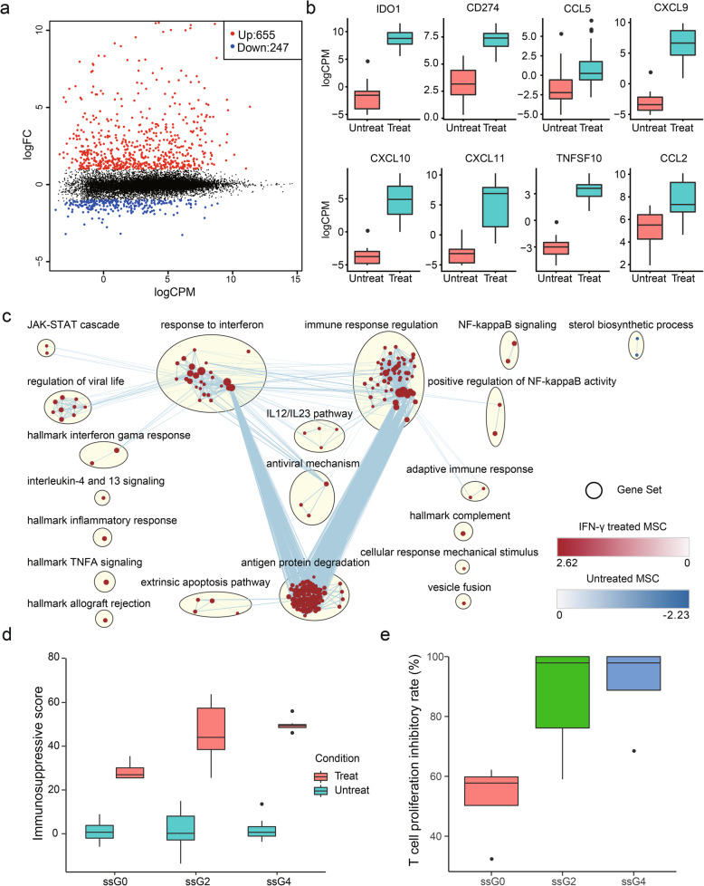
Gene expression changes in MSCs treated with IFNγ. a A mean difference plot showing DEGs identified in MSCs treated with IFNγ. b Representative genes upregulated in IFNγ-licensed MSCs. c Enrichment map showing pathways enriched in INFγ-treated MSCs (red) and untreated MSCs (blue). d Boxplot showing immunosuppressive scores for each group calculated based on VEGF, IFNa, CXCL10, GCSF, CXCL9, IL-7, and CCL2 expression level. e Results showing MSCs from ssG0, ssG2, and ssG4 co-cultured with PBMC for immunosuppressive potency assessment

