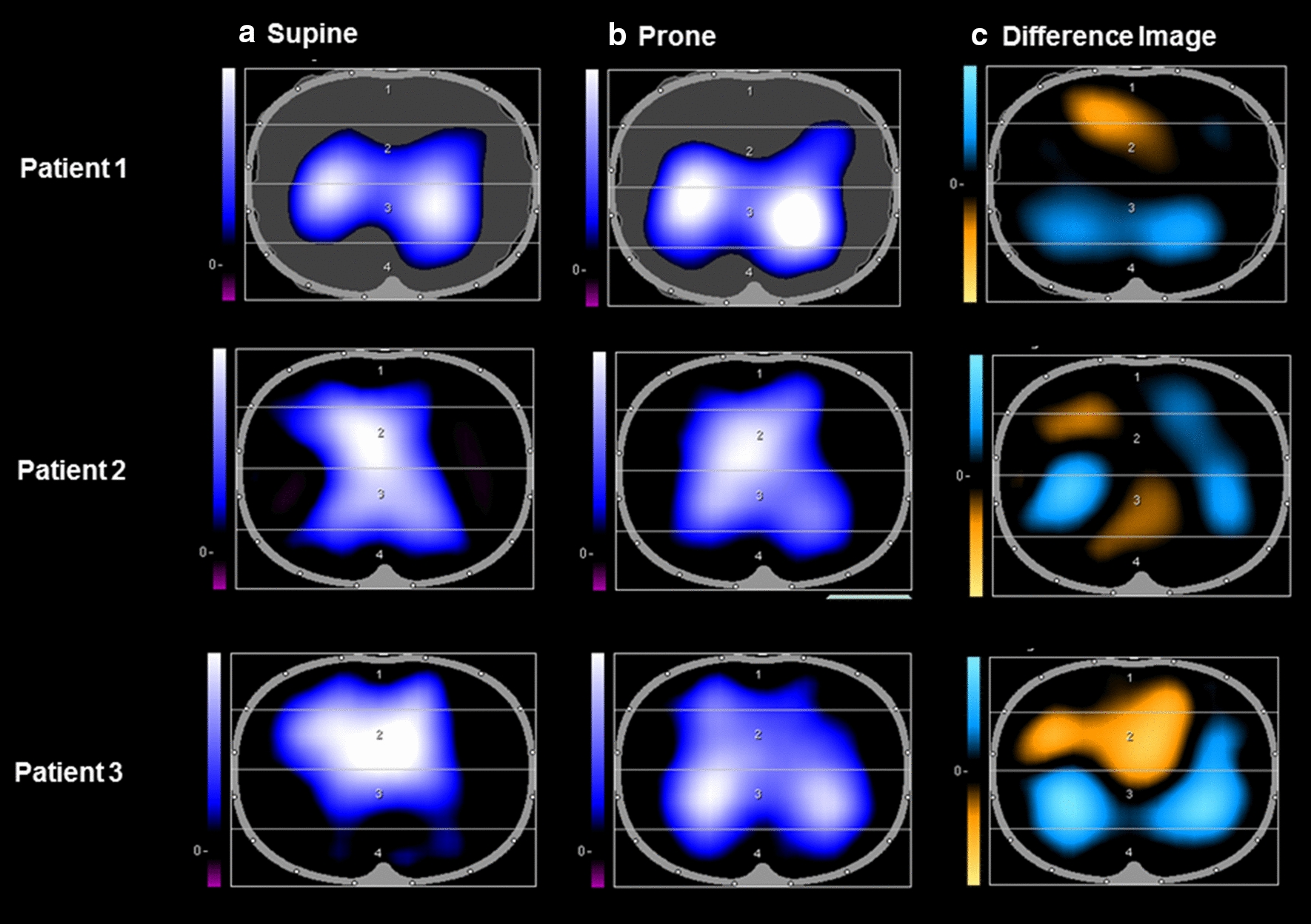
Fig. 2.

Electrical impedance tomographs (PulmoVista 500, Dräger) are shown above for 3 adult patients with SARS-CoV-2 ARDS who were invasively ventilated and underwent prone positioning. a represents the end-inspiratory trend view prior to prone positioning. b represents the end-inspiratory trend view following prone positioning. In (a, b), areas of increasing impedance variation (corresponding to greater ventilation) are represented in order of increasing variation in black (none), blue (intermediate) and white (greatest) colors. c represents the difference between the images in a, b, displaying loss of regional ventilation (areas in orange) which represent ventral regions being over-distended in the supine position and gain of regional ventilation (areas in blue) which represent recruitment of the dorsal regions upon prone positioning. Patients 1 and 3 showed an increase in tidal impedance variation in dorsal regions in the prone position and a decrease in tidal impedance variation in the ventral regions, which is consistent with lung recruitment dorsally.
