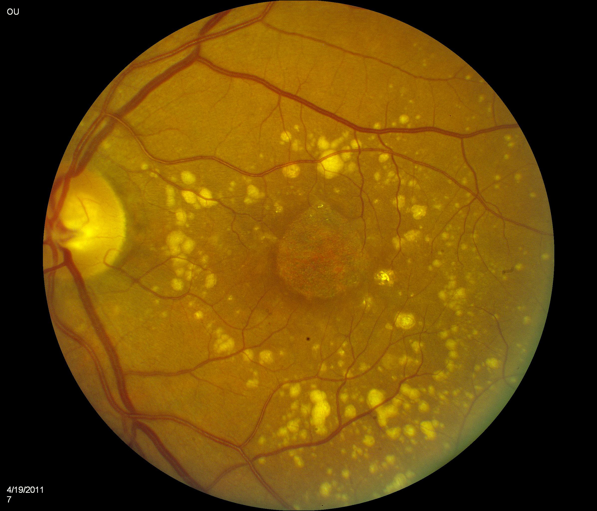
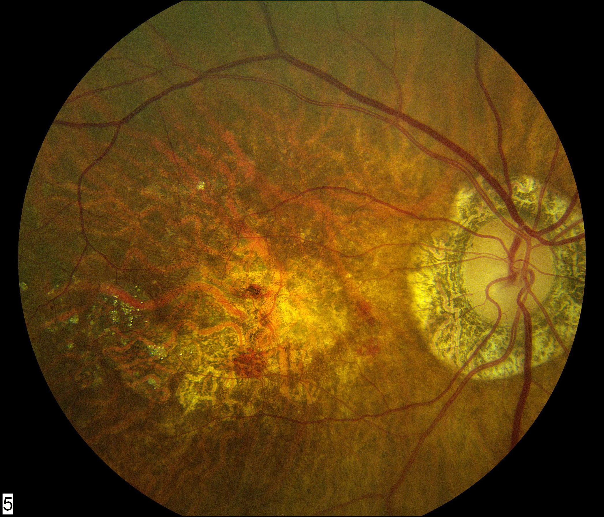
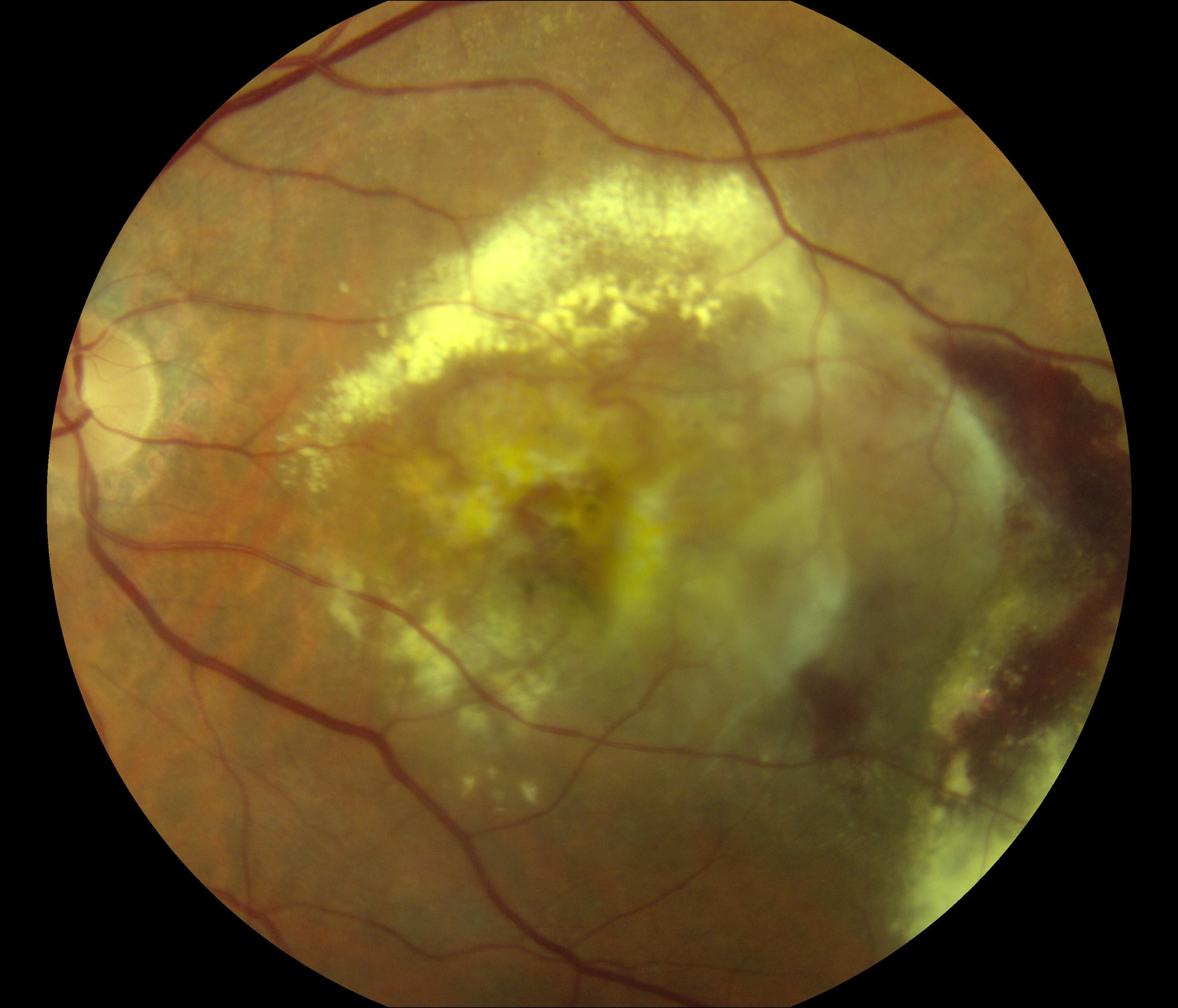
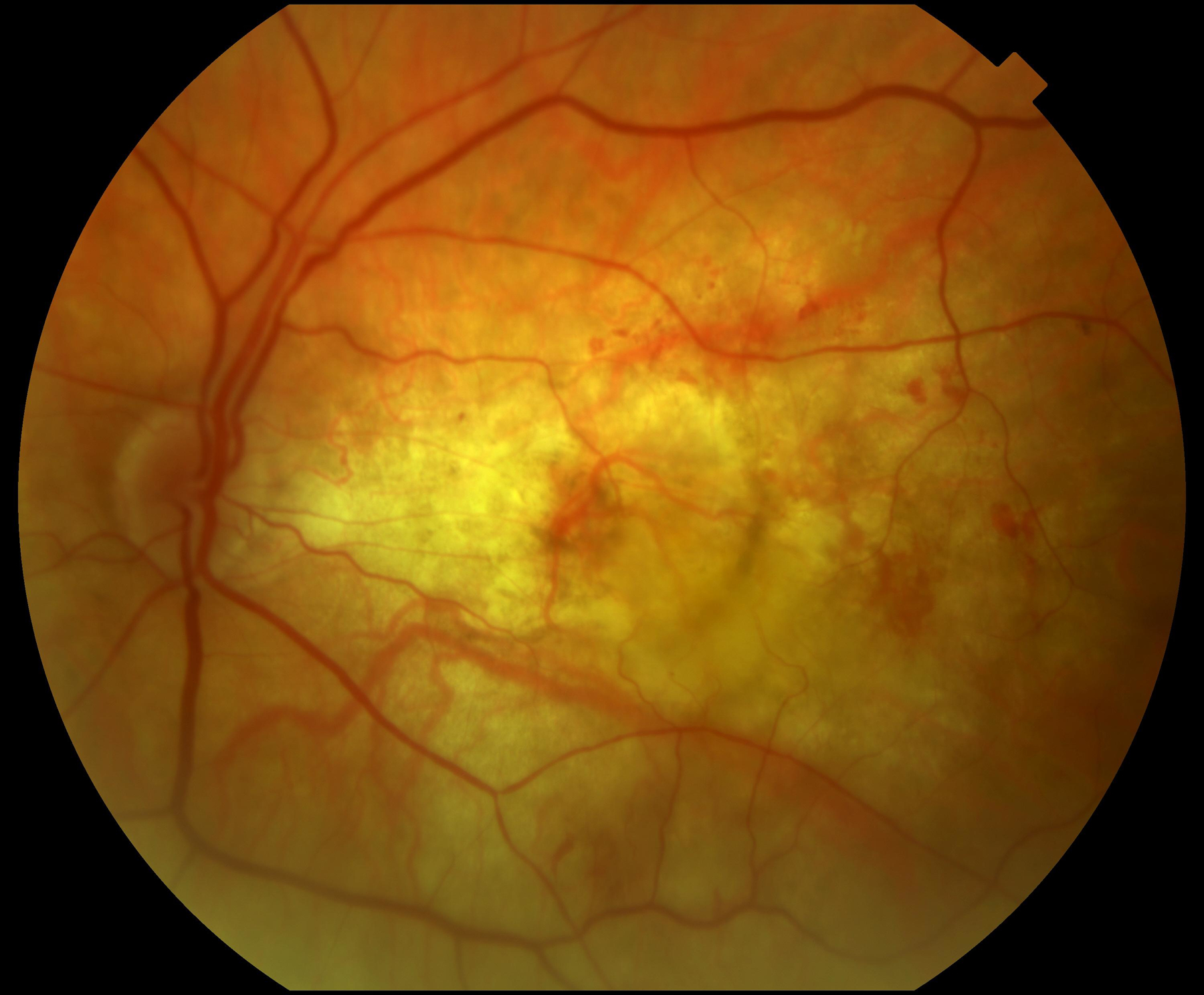
Figure 1.


Color fundus photographs of eyes with refracted best-corrected visual acuity below 20/200 at two years after the first study visit where neovascular age-related macular degeneration was recorded. In (A) and (B), the principal cause for poor acuity was central macular atrophy (without accompanying central subretinal fibrosis). In (C) and (D), the principal cause was central subretinal fibrosis.
