Fig. 1.
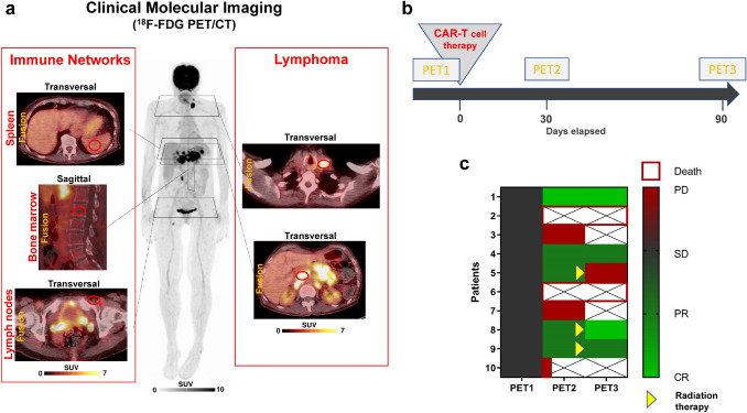
Clinical molecular imaging of the activity of off-target lymphoid organs and lymphoma burden. a Metabolic parameters of lymphoid organs and bone marrow (left panel) and lymphoma burden (right panel) were determined using serial 18F-FDG PET/CT. b Graphical illustration of the time points for serial PET/CT. Patients underwent a baseline scan (PET1) before CAR-T-cell therapy, and early (PET2, + 30d) and late (PET3, + 90d) response assessment scans thereafter, also allowing for assessment of the evolution of metabolic activity. c Graphical illustration of response to CAR-T-cell therapy in DLBCL patients, depicted as heat map ranging from complete remission (light green) to progressive disease (red). Patients continued to receive serial PET if still alive and not showing clear evidence of progression on clinical examination or other imaging (crossed out boxes = PET not performed). Remission at Day 90 required early metabolic response at PET2 in all cases (Fisher’s exact test, P = 0.0476). Patients no. 5, 8 and 9 received consolidation radiation therapy after PET2

