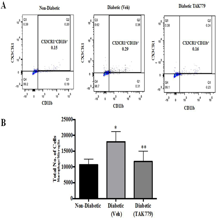
Fig. 3. TAK-779 treatment significantly reduces the infiltration of CX3CR1+/CD11b+ macrophage/microglia cells.
(A) Representative dot plots of flow cytometry analysis of control non-diabetic, diabetic (vehicle) and diabetic (drug treated) STZ mice. Gating was done to include macrophage/microglia cells labelled CX3CR1+/CD11b+. (B) Histogram represents the total number of CX3CR1+/CD11b+ cells, with significantly increased cells in the retinas of diabetic mice (vehicle) in comparison with non-diabetic mice (*p=0.05) and there was a significant decrease in the number of cells in the drug treated animals in comparison with vehicle treated diabetic animals (**p=0.02). Data are mean±S.D.

