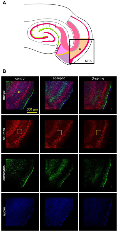
Fig 3: TLE-related pathology in the MEA.
A, Schematic of the MEA-hippocampal brain slice showing the area imaged in B using high resolution confocal microscopy.
B, Immunofluorescence images of the MEA (*, top down panels) in a non-status control (left), epileptic (middle), and post-status rat treated with D-serine (right). Triple immunostaining of neurons (red), astrocytes (green) and nuclei (blue) highlighting neuronal and astroglial pathology within MEA (topmost horizontal panel). Neurons immunoassayed with fluorescently tagged antibodies against NeuN (red, second horizontal panel from top), astrocytes with antibodies against GFAP (green, third horizontal panel from top), and nuclei with DAPI (blue, fourth horizontal panel from top) shown separately [for quantification of data in MEA see (Beesley, et al., 2020)]. The core of the pathology, including cell loss and astrogliosis occurs in layer III of the MEA (rectangular boxes in yellow).

