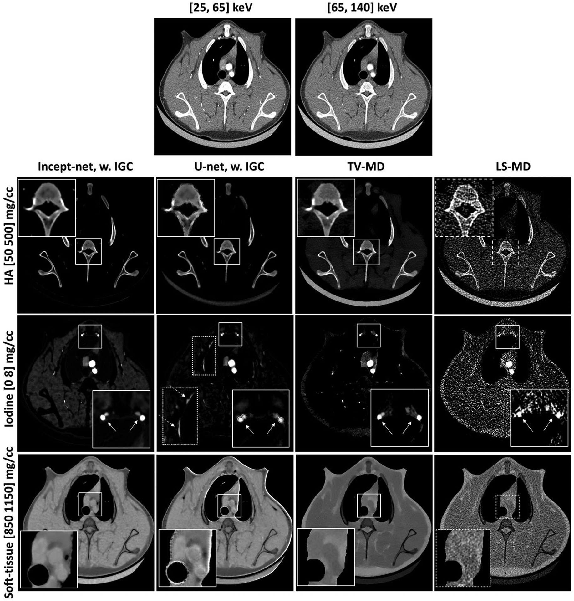
Figure 10.

Material-specific images from a porcine chest CT scan (at a randomly-selected slice), using Incept-net, U-net, total-variation (TV-MD), and least-square (LS-MD) based material decomposition methods. The spatial distribution of hydroxyapatite (HA), iodine and soft-tissue are shown in the top, middle and bottom rows, respectively. The CTDIvol was 16 mGy. The zoomed insets are 2.2 times the size of the square region-of-interests. In the zoomed iodine insets, the solid arrows indicate the location of blood vessels. In the iodine map of U-net outputs, the dashed arrow indicates strong bone residual at chest wall. The range of display window for CT images were [-160, 240] HU.
