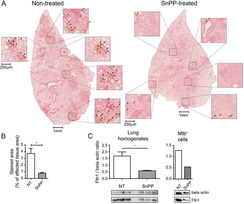
Figure 7. Pharmacological HO-1 inhibition results in decreased iron accumulation in lungs of Mtb-infected mice.
(A) Photomicrographs of whole lung lobes (1 mm scale bars) and magnified fields (250 μm scale bars) showing Perls-DAB staining in histological sections from lungs of Mtb-infected C57BL/6 mice that were treated or not with SnPP for 21 days starting at 28 days post-infection (dpi) – arrowheads indicate areas with positive Perls-DAB staining; (B) Graphs showing percentage represented by Perls-DAB positively stained area in regions containing infiltration of inflammatory cells, in histological sections from lungs of Mtb-infected C57BL/6 mice treated or not SnPP for 21 days starting at 28 dpi (n = 4 and 3 mice/group); (C) Graphs showing quantification of ferritin heavy chain (Fth1) expression normalized to that of beta actin (endogenous control) (top) and images of western blot membrane showing Fth1 and beta actin expression (bottom) in lung homogenates (left) and mCherry Mtb+ pulmonary leukocytes isolated by sorting (right) from C57BL/6 Mtb-infected mice treated or not with SnPP for 21 days starting at 28 days post-infection (n = 3 mice/group on left panel and pooled samples from 3 mice/group in right panel). The data are presented as the means ± standard error (B and C), photomicrographs of individual samples (A) or individual samples (C). Data shown are representative of 2 independent experiments. Statistical analysis: Student’s t test. * = p<0.05.

