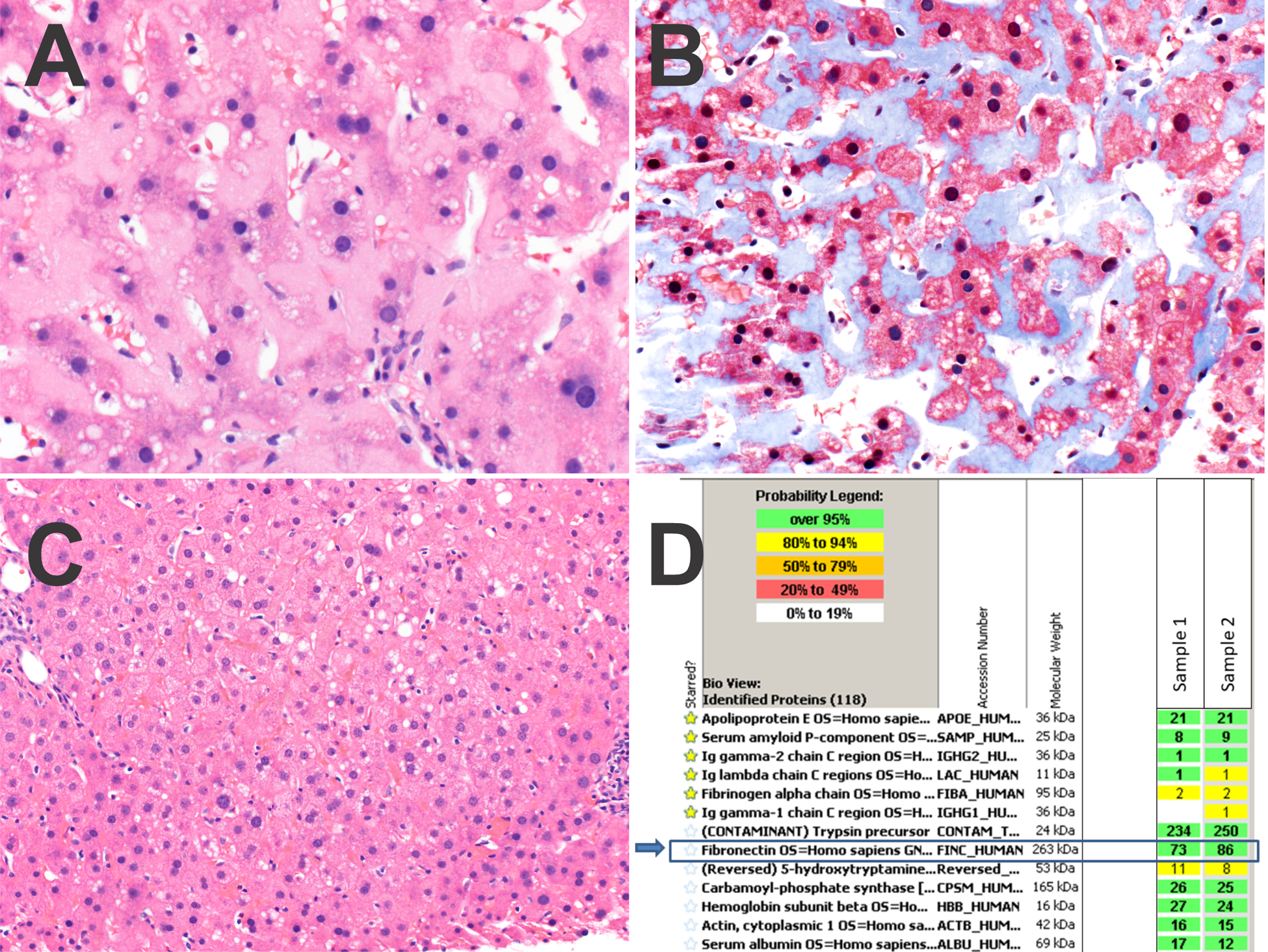
Figure 2. Case 2.

Panel A. The sinusoids show dense deposits of fibronectin.
Panel B. A trichrome stain stains the fibronectin light blue.
Panel C. The fibronectin deposition was patchy, with other areas of the biopsy negative for fibronectin.
Panel D. Protein identification reports from LC-MS/MS analysis. The extracellular deposits were microdissected in duplicate (sample 1 and 2). The starred amyloid-associated proteins are not abundant. Rather, fibronectin is the dominant protein in the deposits (blue arrows). Trypsin is present as it is the enzyme used in digestion of the peptides prior to analysis.
