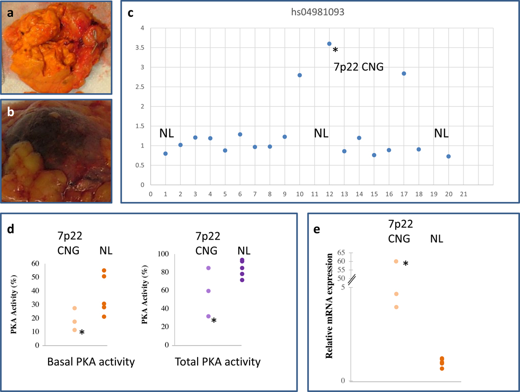
Fig. 3. PKA activity and PRKAR1B subunit expression in tumors with 7p22 CNG.
(a) Tumor sample ADT35.02. (b) Tumor sample ADT47.03. (c) Detection of 7p22 CNG, by the hs04981093 probe, in tumors ADT35.02, ADT47.03 and ADT183.02 at positions 10, 12 and 17 respectively. (d) PKA activity was measured in tumor cell lysates. The 3 tumors with 7p22 CNG had lower basal PKA activity (in absence of cAMP, left panel) but no differences in total PKA activity (in presence of cAMP, right panel) from NL CPA (N=5) that did not contain 7p22 CNG. (e) The 3 CPA with 7p22 CNG showed higher expression of the PRKAR1B mRNA compared to the 5 CPA that were genomically NL. NL stands for Normal and the asterisk (*) corresponds to tumor ADT47.03 as this is the CPA that also carried the L206R PRKACA “hot-spot” pathogenic variant.

