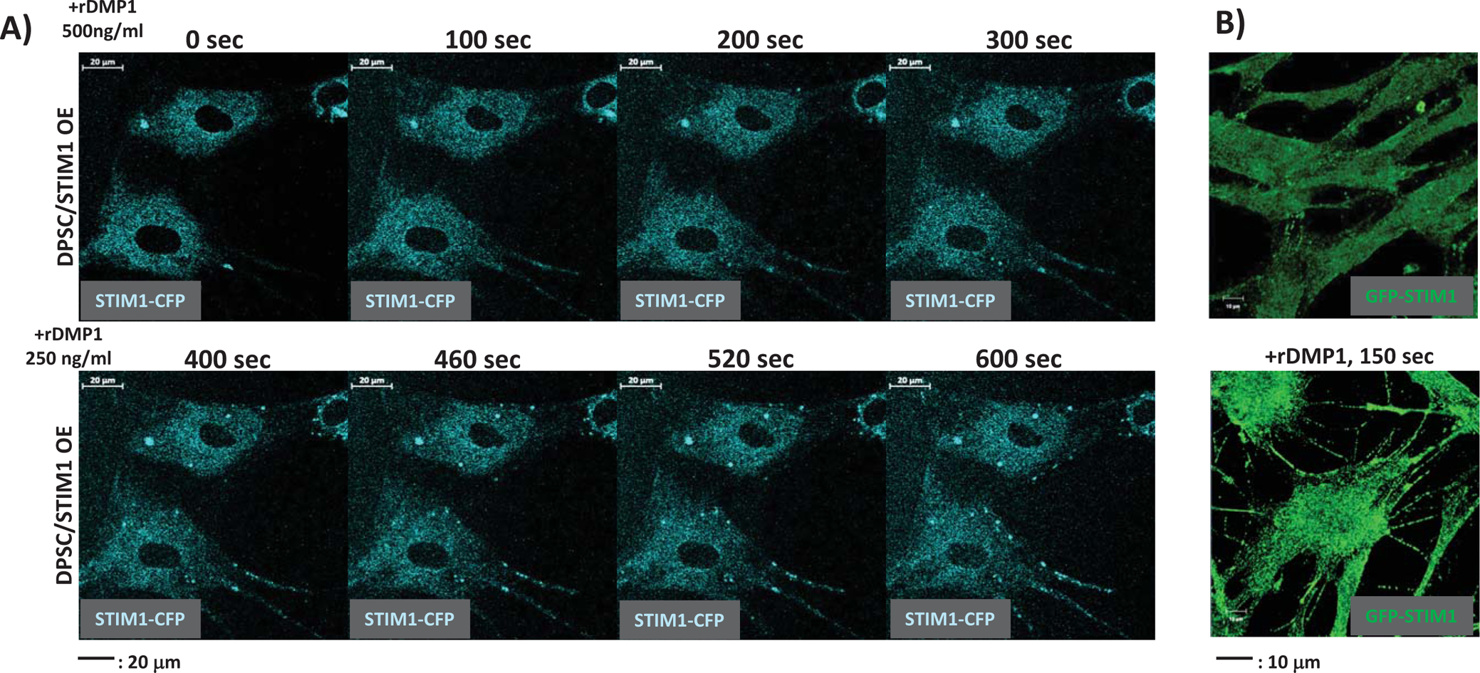
Fig 3. Time course of spatial mobilization and puncta formation of STIM1 with ER Ca2+ depletion.

(A) Dental pulp stem cells transfected with CFP-STIM1 were stimulated by 500 ng/ml DMP1 for various time periods to trigger ER Ca2+ release. Oligomerization and mobilization of STIM1 were monitored by live-cell imaging using confocal microscopy. Note “puncta” formation with ER store depletion.
(B) Changes in cell morphology of DPSC–GFP-STIM1 overexpressing cells with ER Ca2+ release triggered by the addition of DMP1. Note formation of extensive cellular process.
