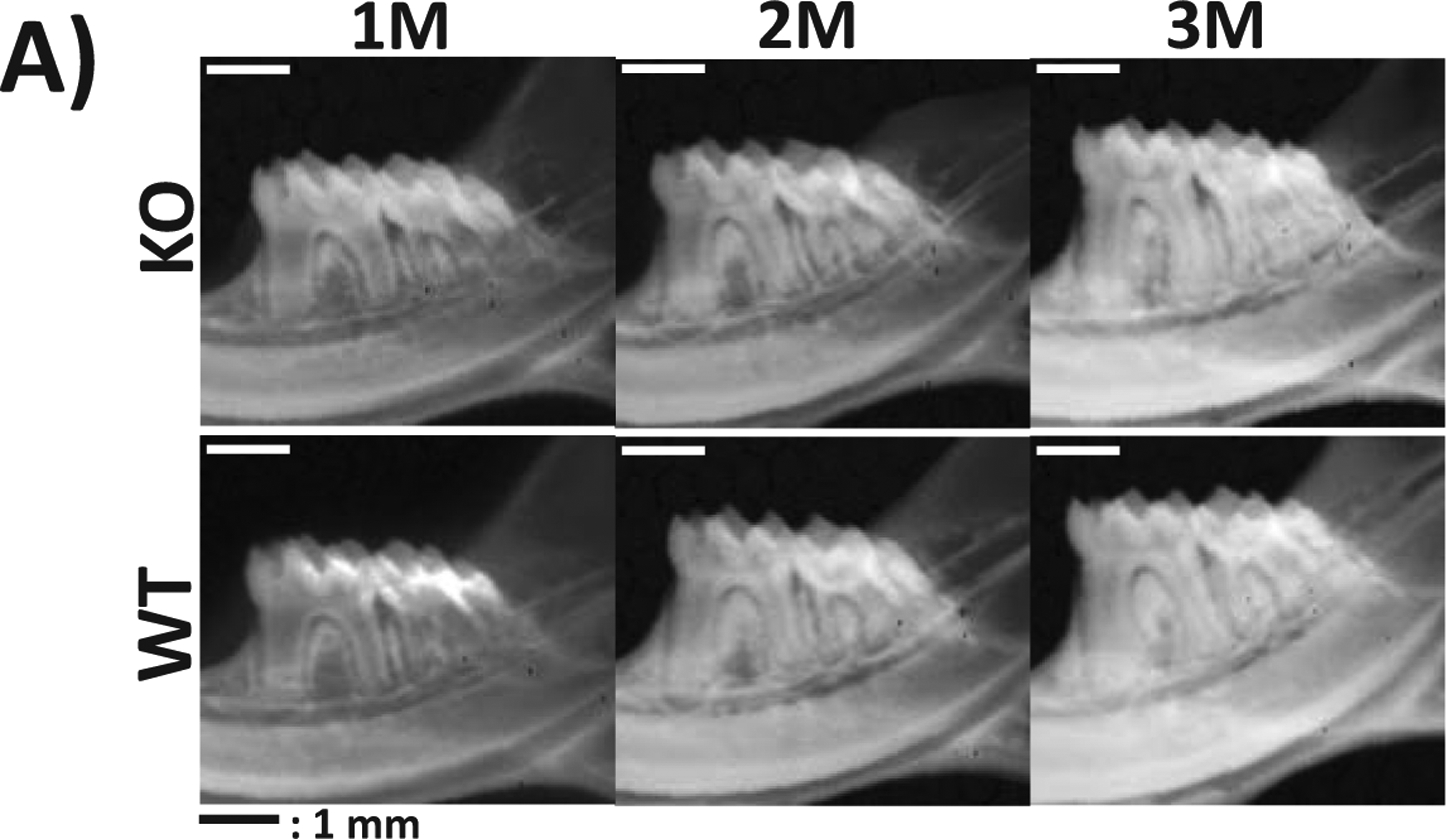
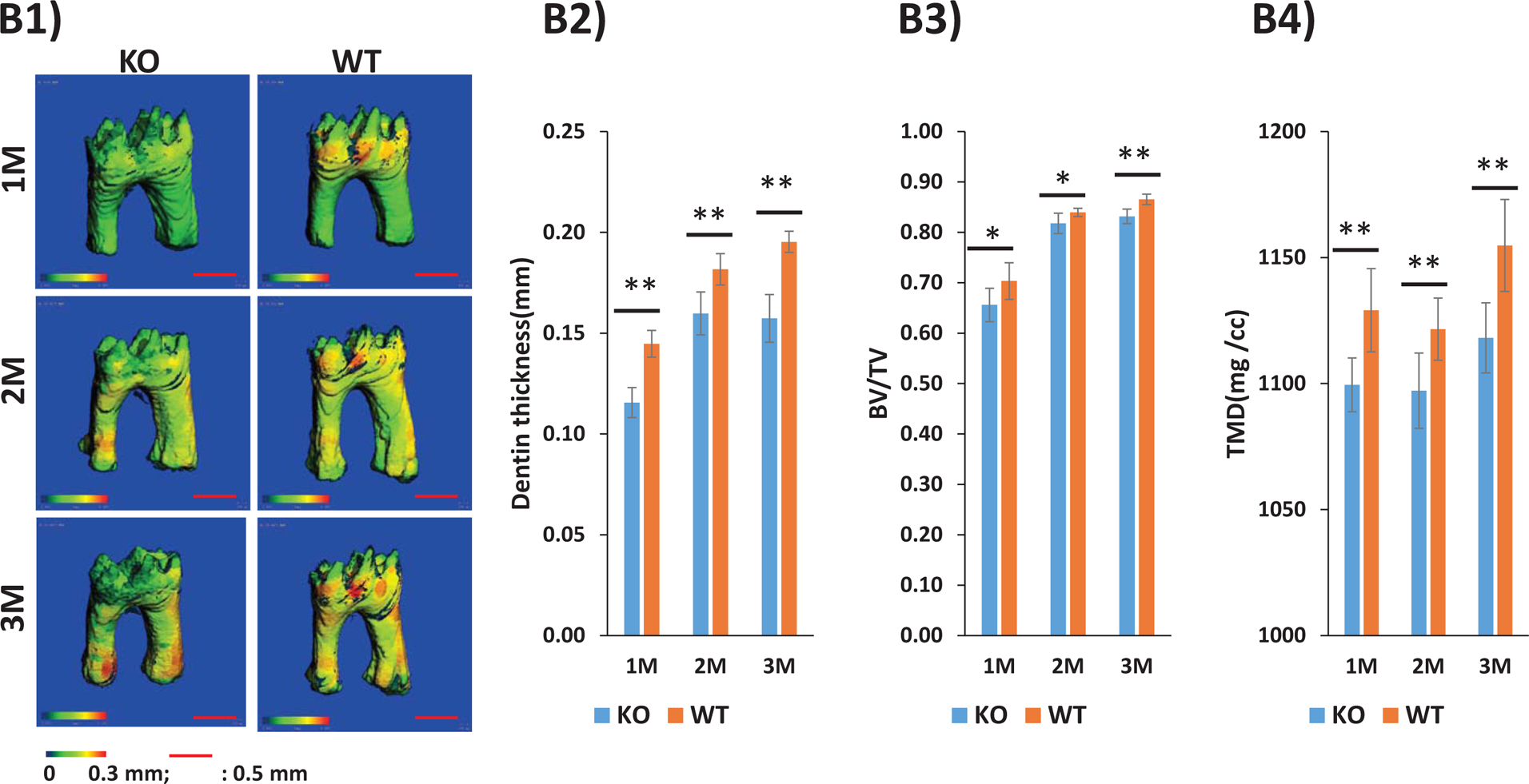
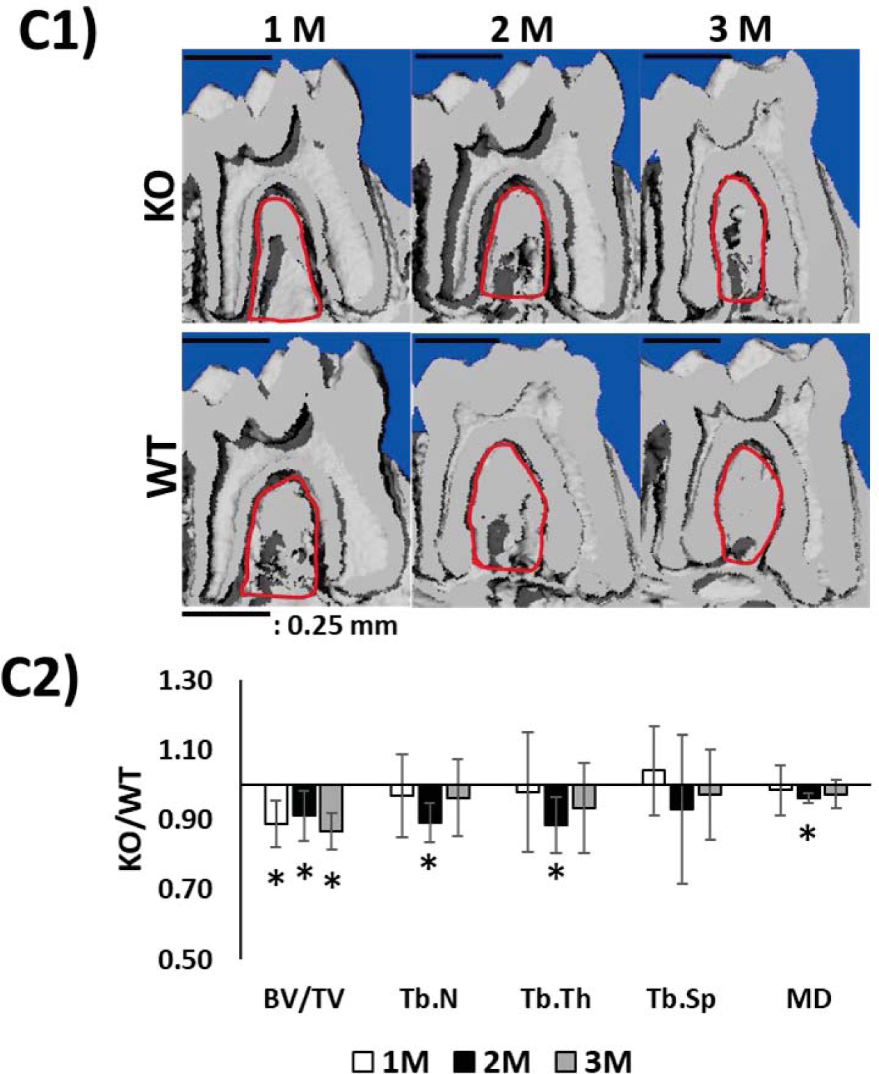
Fig 7. Characterization of the tooth-alveolar bone complex of STIM1 knockout mice by radiographic and micro-computed tomographic analyses:


(A) Representative radiographs show disordered alveolar bone and dentin at 1, 2 and 3 months of age in STIM1-KO mice when compared with the wild type (WT).
(B) Representative segmentation analysis of the micro-CT obtained images of the first molar from 1, 2 & 3 months WT and STIM1-KO mice (n=6). Overall dentin thickness varies from 0–0.3mm based on segmentation analysis (B1). Dentin thickness, Dentin volume fraction (BV/TV) and tissue mineral density (TMD) are shown in B2–B4 respectively.
(C) The μCT 3D longitudinal segmentations showing alveolar bone defects surrounding the first molar (C1, red circles). (C2) shows morphometric comparison (KO/WT) of the alveolar bone. Note decrease in bone volume (BV/TV); Trabecular number Tb.N, trabecular thickness Tb.Th, trabecular spacing Tb.Sp and mineral density MD. Statistical significance: *: p< 0.05
