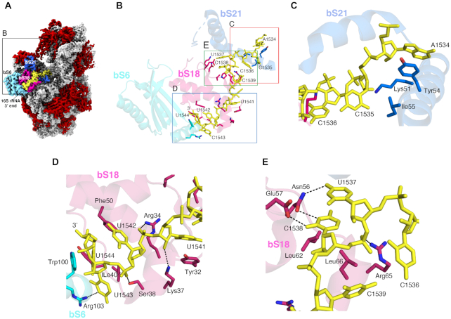
Figure 5.
Structural Basis for the Sequestration of the ASD sequence within the F. johnsoniae 30S subunit. (A) Solvent view of the cryo-EM structure of the F. johnsoniae 30S subunit showing the 3′ end segment of 16S rRNA (highlighted in yellow). The remaining 16S rRNA is shown in light grey. The r proteins bS6, bS18 and bS21 involved in the sequestration of the SD sequence are colored in cyan, pink and blue, respectively. The remaining r proteins are shown in red. Zoomed area shown in (B) is framed in the structure. (B) Interaction details between the 3′ end nucleotides (including the ASD sequence) of 16S rRNA (yellow sticks) and r proteins bS6 (cyan), bS18 (pink), and bS21 (dark blue). Zoomed areas shown in (C), (D) and (E) are indicated in panel (B).

