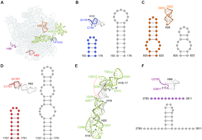
Figure 7.
Comparison of structural differences in the 23S rRNA between the 50S subunit in F. johnsoniae and E. coli. (A) Solvent view of the 23S rRNA from F. johnsoniae with the regions distinct from E. coli (PDB ID: 6O9K) labelled and highlighted in different colors. (B–F) Comparison of helices H10, H28, H63, H16-H17, and H98, respectively from F. johnsoniae and E. coli along with their tertiary structure derived secondary structure diagram (except for helices H16-H17). Color codes for F. johnsoniae 23S rRNA elements are same as in (A), and E. coli’s elements are shown in grey. The red dashed lines in (E) indicate the segment which could not be modeled due to weaker densities. Comparisons with T. thermophilus, M. smegmatis, P. aeruginosa, B. subtilis, and S. aureus are shown in Supplementary Figure S16.

