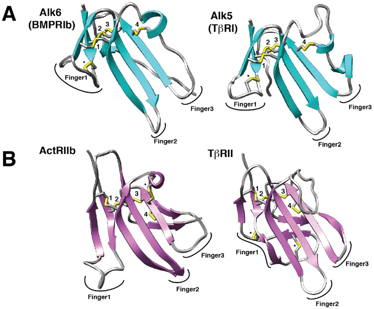
Figure 3.
Structures of representative type I and type II receptors of the TGF-β family. A, B. Structures of type I and type II receptors (A and B, respectively) shown as ribbon diagrams. Disulfide bonds are depicted as sticks with yellow sulfur atoms. Four disulfide bonds that are positionally conserved in all type I and type II receptors of the TGF-β family are labeled 1 – 4. Disulfide bonds that are unique are labeled with an asterisk (*). The three fingers of the type I and type II receptors that define them as having a three-finger toxin fold are indicated. Structures shown correspond to the following PDB entries: Alk6 – 3EVS 103, Alk5 – 2L5S 104, ActRIIb – 1NYU 105, and TβRII – 1M9Z 106.

