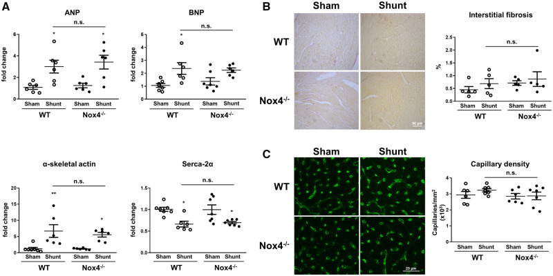
Figure 4.
Cardiac gene expression, fibrosis, and capillary density in Nox4−/− mice and WT littermates after volume overload. (A) LV mRNA levels of ANP (atrial natriuretic peptide), BNP (brain natriuretic peptide), α-skeletal actin, and SERCA-2α (sarcoplasmic/endoplasmic reticulum calcium ATPase-2α) after Shunt or Sham control. (B and C) Representative images and mean data of Picrosirius red-stained sections indicating fibrotic regions (B) and isolectin B4-staining depicting cardiac capillaries (C) after Shunt or Sham (n = 5–7/group). *P < 0.05, **P < 0.01 in Shunt vs. Sham, n.s., not significant between genotypes using two-way ANOVA followed by Bonferroni post-hoc test for multiple comparisons.

