Figure 6.
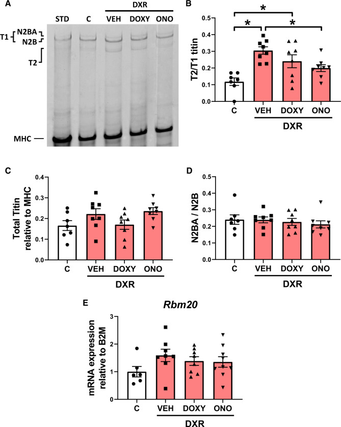
Cardiac titin proteolysis and isoform expression in DXR cardiotoxicity. (A) Representative Coomassie blue-stained agarose gel showing titin levels in ventricular extracts. The ratio of titin degradation product (T2) to N2BA and N2B titin (T1) was determined in left ventricular extracts from control, DXR, DXR + Doxy, and DXR + ONO groups. MHC was used as a loading control. (B) Cardiac titin proteolysis, represented by the T2 to T1 titin ratio, was significantly increased in DXR and DXR + Doxy mice. DXR-induced titin proteolysis was prevented with ONO (n = 7–8). (C) Ratio of total titin (T1 + T2) to MHC content was unchanged between groups (n = 7–8). (D and E) DXR did not alter cardiac titin isoform expression, seen by the absence of changes in N2BA to N2B titin (n = 7–8) and Rbm20 mRNA expression (n = 6–8). STD is a standard ventricular extract from a control mouse that was not part of this study. *P < 0.05 by one-way ANOVA followed by the Sidak’s post hoc test.

