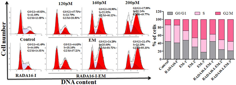
Figure 6.
Hepa1-6 cells incubated with free EM (water suspension) or RADA16-I-EM hydrogels for 24 h and the cell cycles analyzed under flow cytometry. The percentage of G0/G1, S and G2/M phase distributions of free EM or RADA16-I-EM hydrogels treated cells was measured by ModFit. LT 3.3 software. 1, 2 and 3 represent EM concentrations of 120, 160, 200 μM, respectively, [RADA16-I] = 5 mg/mL.

