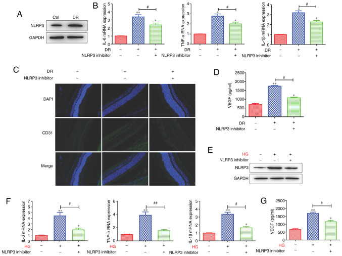
Figure 2.
NLRP3-mediated tissue inflammatory response promotes microvascular cell proliferation in the retina. (A) The protein expression level of NLRP3 was increased in the DR groups. (B) Following the of blocking NLRP3, the mRNA expression of IL-6, TNF-α and IL-1β was downregulated. (C) The expression level of the vascular marker, CD31, and (D) the VEGF secretion level were decreased following the inhibition of NLRP3. In addition, in the HRMEC cell model, (E) HG enhanced the protein expression of NLRP3, which was inhibited by NLPR3 inhibitor. (F) The levels of the inflammatory-related cytokines, IL-6, TNF-α and IL-1β, were enhanced in the HG-induced HRMEC cell model and were inhibited by the blocking of NLRP3. (G) The VEGF secretion level was enhanced, and was decreased following the inhibition of NLRP3. Data are presented as the mean ± standard deviation from triplicate wells. *P<0.05 and **P<0.01 compared with the control; #P<0.05 and ##P<0.01 compared with the relative DR animal model group or HG-induced HRMEC cell group. NLRP3, NLR family pyrin domain containing 3; DR, diabetic retinopathy; IL, interleukin; TNF, tumor necrosis factor; VEGF, vascular endothelial growth factor; HG, high glucose; HRMEC, human retinal microvascular endothelial cell; Ctrl, control.

