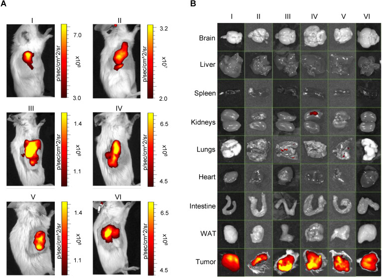
Figure 4.
Pseudocolor representation of ICG fluorescence acquired by in vivo (A, left panels) and related ex vivo (B, right panels) imaging of isolated organs of n=6 PDX mice (I, II, III, IV, V, and VI) injected with EVs derived from the plasma of the same patients from which the corresponding PDX model was generated. In all cases, tumors were selectively labeled with ICG-loaded EVs. WAT: white adipose tissue.

