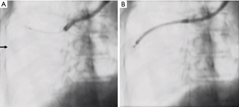
Figure 2.
Fluoroscopic images showing the accessibility of a PPL using a 3.0-mm ultrathin bronchoscope and a 4.0-mm bronchoscope. (A) The EBUS probe could not be advanced towards the target lesion (arrow) using a 4.0-mm bronchoscope. (B) The 3.0-mm ultrathin bronchoscope approached the lesion and provided a diagnosis of adenocarcinoma. PPL, peripheral pulmonary lesion; EBUS, endobronchial ultrasound.

